Background: Venous return from the lower extremities is pumped upwards to the right side of the heart in a process that is facilitated by one-way valves and the venous muscle pump, of which the calf muscle pump is a major contributor1-3. Venous plethysmography can quantitatively assess calf pump function (CPF). The association between the CPF and venous thromboembolism (VTE) has not been investigated.
Methods: Venous plethysmography (VP) data (strain gauge or air plethysmography) from the Mayo Clinic Vascular Lab database (1998-2015) of CPF (bilaterally reduced, unilaterally reduced, and bilateral normal) were examined in Olmsted County Residents. The Rochester Epidemiology Project (REP) captures the population of Olmsted County and contains demographic information, medical diagnoses, hospital admissions, and surgical procedures as well as validated VTE events and death. Patients with signs of obstructed outflow in either extremity on the venous plethysmography (a possible sign of acute or chronic deep vein thrombosis) study were excluded. Patients with a history of VTE diagnosed before the physiologic study were also excluded. If more than one measurement of calf muscle pump function was performed, only the first measurement was used. The primary outcome was a composite of any VTE, including proximal and distal deep vein thrombosis (DVT) and pulmonary embolism (PE).
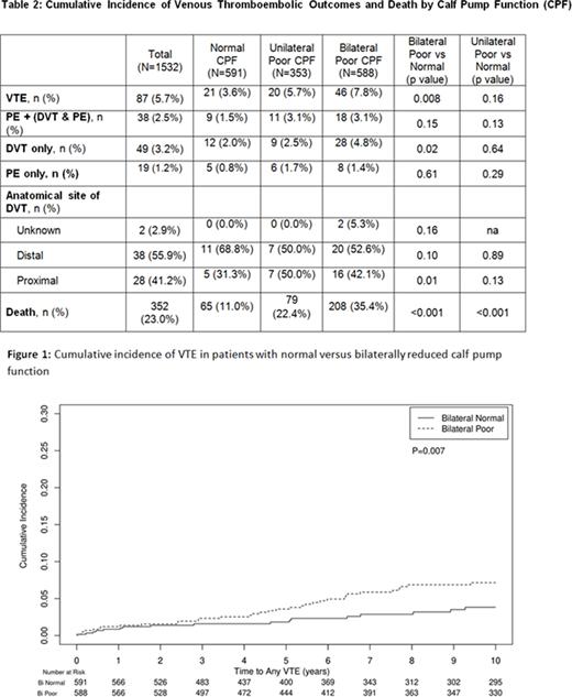
Results: 1703 Olmsted County residents had venous plethysmography studies performed. MN research authorization was denied in 64 patients and 107 were excluded for any documented VTE preceding index VP study. 1532 patients with recorded CPF (28% air and 72% strain gauge plethysmography) were studied: 591 (38.5%) had normal CPF, 353 (23.0%) had unilateral reduced CPF (rCPF), and 588 (38.3%) had bilateral rCPF. The mean age was 64.4 (SD 18.4), 68.9% were female, and the mean BMI was 29.5 (SD 6.4). Any VTE occurred in 87 patients (5.7%) after a mean follow up of 10.9 years (range 0-22.0 years). Isolated lower extremity DVT (excluding concurrent PE) occurred in 49 patients and PE+/-DVT occurred in 38 patients. Death occurred in 352 patients (23%). Bilateral rCPF compared to bilateral normal CPF was associated with VTE (p=0.007), DVT only (p=0.02) and death (p<0.001) but not PE+/-DVT (p=0.13). Unilateral rCPF compared to bilateral normal CPF was not associated with VTE, but was associated with death (p<0.001). Kaplan-Meier curves for VTE and death are shown in Figure 1. The hazard ratio for bilateral rCPF compared to bilateral normal CPF for VTE was 2.0 (95% CI 1.2-3.4) and for DVT only was 2.2 (95% CI 1.1-4.2). A sensitivity analysis for the main outcome of VTE did not show significant interaction based on the type of plethysmography (strain vs. air), by age stratified at 65 years, sex, or BMI stratified at 30 (p>0.1 for each comparison).
Conclusion: In this population-based study of Olmsted County residents with no prior VTE, rCPF function as measured by venous plethysmography is associated with increased risk for VTE, particularly lower extremity proximal DVT. More research is required to understand what additional measures of venous physiology influence these findings and whether CPF could be used in VTE risk stratification.
No relevant conflicts of interest to declare.
Author notes
Asterisk with author names denotes non-ASH members.


This feature is available to Subscribers Only
Sign In or Create an Account Close Modal